
SKELETAL SYSTEM ANALYSIS OF ASIAN GIANT SOFT-SHELLED TURTLES AND COMPARISON WITH CHINESE SOFT-SHELL TURTLE
-
摘要: 采用虫蚀法制备了鼋Pelochelys cantorii和中华鳖Pelodiscus sinensis的骨骼标本, 对骨骼系统进行了观察、描述、绘图及比较分析。结果显示, 鼋的骨骼共169枚, 由背甲和腹甲组成的外骨骼、中轴骨和附肢骨组成的内骨骼组成。通过比较鼋和中华鳖的骨骼结构, 发现二者在头骨的吻突长度及第三颈椎结构方面有较大差别。鼋眼眶前部至吻突最前端的长度与头骨总长度比为0.082, 而中华鳖为0.570, 显示中华鳖吻突显著长于鼋。鼋与中华鳖的颈椎骨数目均为9枚, 但中华鳖的第三至第九颈椎的横突要更明显, 第七颈椎的椎体向上显著翘起, 且第九颈椎腹面椎体前端为尖状。从整条颈椎上看, 鼋脊椎长度与其背甲长度之比为0.66, 中华鳖为1.07, 表明中华鳖的颈椎更长; 研究结果丰富了鳖科动物的骨骼学基础数据, 也为鼋物种鉴定、龟鳖动物系统演化及生态适应性提供骨骼学理论依据。Abstract: Asian giant soft-shelled turtle is one of the first class protected animals in China, and is an extremely endangered species. Now, there are only 13 sexually mature Asian giant soft-shelled turtles in captivity in China. Due to fill the blank in the study of Asian giant soft-shelled turtle skeleton system, this study described the morphological characteristics of the complete skeletal system of Asian giant soft-shelled turtles and Chinese soft-shell turtles, and draws a diagram. The frozen samples of two Asian giant soft-shelled turtles and one Chinese soft-shell turtle were studied in this study. After the processes such asdefrosting and dissection, we used larva of Tenebrio molitor to manufactured skull specimen of the turtles. Removed the bones and rinsed with water to dry them. Then the vernier calipers were used to measured each bone structure and take photos, and the shapes were hand-drawn with reference to the bone entities and photos. A total of 169 bone fragments were found, consisting of exoskeleton of dorsal and ventral armor, endoskeleton of midshaft bone and appendage bone. Meanwhile, the skeletal structure of Asian giant soft-shelled turtle and Chinese soft-shell turtle had significant differences in snout length and the third cervical spine structure. The ratio of the length from the anterior orbit of soft-sheller to the most tip of the muzzle to the total length of skull was 0.082 for Asian giant soft-shelled turtle and 0.570 for the Chinese soft-shell turtle. Although the number of cervical vertebrae bones of Asian giant soft-shelled turtle and Chinese soft-shell turtle was 9, the transverse process of the third to ninth cervical vertebrae of Chinese soft-shell turtle was more obvious, the cone of the seventh cervical vertebrae was obviously warped upward, and the front end of the abdominal cone of the ninth cervical vertebrae was pointed. From the whole cervical spine, the ratio of the length of the spine of to the length of its back armor is 0.66 for the Asian giant soft-shelled turtle and 1.07 for the Chinese soft-shell turtle. These results provide the basis for the identification of Asian giant soft-shelled turtles, the phylogenetic evolution and ecological adaptability of turtles, indicating that Asian giant soft-shelled turtles are primitive reptiles with an important role in animal evolution.
-
骨骼是组成脊椎动物内骨骼的坚硬器官, 作为生物个体运动的基础, 起着增大生物体体积、加速器官进化、支撑和保护动物的作用, 在生物协同进化的过程中也扮演了重要角色[1], 在动物的进化过程中, 各个物种为了适应不同的生存环境而出现了分化, 其骨骼也发生相应改变, 例如鲸类的四肢演化成鳍状, 四肢长骨也相应变得粗短[2]。所以, 研究脊椎动物骨骼的形态区别, 有利于从骨骼进化方面寻找物种分类的依据, 也便于解释不同种属生物的行为差异。同时, 受加工的影响, 常常难以在骨骼上取得足够鉴定所需DNA量, 此时骨骼形态学就可作为一种鉴定物种的有效方式, 可以用于打击濒危动物非法贸易。
龟鳖目曾经是一个物种多样性非常丰富的类群, 发现最早的龟鳖目动物化石大约出现在2亿年前, 但如今仅存在15科96属300多种[3], 6科21属41种[4]。骨骼形态学也运用在龟鳖目动物的研究上, Caffney[5]对澳大利亚和南美产的蛇颈龟科(Chelidae)的7个属进行了头骨解剖, 依据头骨的形状用分支系统学方法重新对蛇颈龟科的系统发育关系和分类进行了修订。Melan[6]根据113个骨学性状对鳖科(Trionychidae)进行分支系统学研究, 为鳖科内种类的共同祖先和分支进行了探讨。国内学者傅金钟[7]基于骨骼系统的差异对中国龟类进行了分类学研究, 对闭壳龟属和盒龟属的差异做了简单总结。此外, 国内对于龟鳖类骨骼系统较为完整的见刊文章仅有中华鳖(Pelodiscus sinensis)[8]和四爪陆龟(Testudo horsfieldii)[9]。
鼋(Pelochelys cantorii)隶属龟鳖目(Testudines)、鳖科(Trionychidae)、鼋属(Pelochelys), 是我国内陆体型最大的水生龟鳖动物之一, 其种族延续历史悠久, 在古地理研究和古生物进化等方面具有重要的科学价值。鼋在历史上广泛分布于我国东南亚地区, 但由于不正当的贸易、人类的滥捕滥杀及水利工程等等, 导致生境不断恶化, 鼋种群数量锐减[10], 现如今, 鼋已经处于极度濒危状态, 仅在人工条件下保有性成熟鼋13只[11, 12]。我国1989年将鼋列为国家一级重点水生野生保护动物, 世界自然保护联盟(IUCN)在2000年认定鼋为濒危动物, 濒临绝种野生动植物国际贸易公约(CITES)在2003年将鼋列于附录Ⅱ名单。然而, 目前针对鼋的研究主要集中在繁育方式探究和种质资源调查上[11], 对于鼋骨骼系统的研究仅见于《中国动物志·爬行纲》中一些简单的文字描述[13]。总之, 现有的鼋骨骼系统资料匮乏, 尚不能满足开展其基础生物学研究的需要。本研究对鼋骨骼系统进行研究, 为鼋在龟鳖动物分类学及生态适应性等方面的研究提供资料。
1. 材料与方法
1.1 实验对象
用于骨骼系统研究的鼋个体为两只人工繁育的子一代个体, 因病死亡后–20℃冻存。其中一只鼋的背甲长7.48 cm(标本号为ZJ-20211211), 另一只鼋的背甲长10.51 cm(标号为ZJ-20211228)。中华鳖样是人工养殖的商业鳖(编号为TS-20211228)。
1.2 动物CT
利用DELTA计算机断层扫描仪器DELab μCT-100(中国台湾)对冰冻的鼋及中华鳖进行临床前小动物Micro- CT技术, 做X-ray扫描。
1.3 骨骼标本制作
采用虫蚀法, 用黄粉虫(Tenebrio molitor)进行骨骼标本制作。将保存的鼋样本取出, 在室温下解冻后, 用解剖剪将鼋和中华鳖的背甲与腹甲连接处剪开, 沿腹甲边缘剪下整个腹部, 可见整个体腔, 用镊子将内脏全部取出, 刮除大块的脂肪。然后将处理好的样本置于65℃水浴中30min。捞出后, 吸干样本上的水分, 放于盒中冷却, 倒入适量黄粉虫, 黄粉虫的数量以能包覆整个样本躯体为佳, 根据黄粉虫的最佳培养温度及湿度, 将整个盒置于25℃孵化箱中。体长较小的鼋(ZJ-20211211)虫蚀处理过程约持续20h, 体长较长的鼋(ZJ-20211228)和中华鳖(TS-20211228)约经黄粉虫处理40h即可。
1.4 形态测绘
取出骨骼并用清水冲洗、擦干、拍照, 再用游标卡尺(0.01 cm)对各骨骼结构进行测量, 参照骨骼实体和照片对其形态进行手绘(根据骨骼制作完整情况, 鼋背甲腹面及头骨绘自ZJ-20211211, 其余结构以鼋ZJ-20211228骨骼标本为绘图模板), 其骨骼命名参考中华鳖骨骼系统[8]及四爪陆龟骨骼系统[9]的研究。
1.5 漂白及脱脂
骨骼带有较多油脂, 必须进行脱脂以长期保存。本实验采用生活中常用的洗洁精脱去油脂。将腐蚀好的骨骼用清水洗干净并晾干, 浸入10%洗洁精水溶液中浸泡12h。将脱脂后的骨骼先放入清水中洗干净并晾干, 然后放入5%H2O2溶液中浸泡2—7h, 不同骨骼漂白所需要的时间不同。
2. 结果
2.1 鼋的骨骼系统
鼋的骨骼系统总述 观察得鼋的骨骼共169枚(紧密愈合的骨块视作一枚骨)。整个骨骼系统由外骨骼和内骨骼组成, 背甲(Carapace)和腹甲(Plastron)为外骨骼; 脊柱(Spine)和头骨(Skull)等中轴骨; 肩带(Pectoral girdle)、前肢骨(Forelimb skeleton)、腰带(Pelvic girdle)和后肢骨(Pelvic appendage)等附肢骨均为内骨骼(图 1和图 2)。
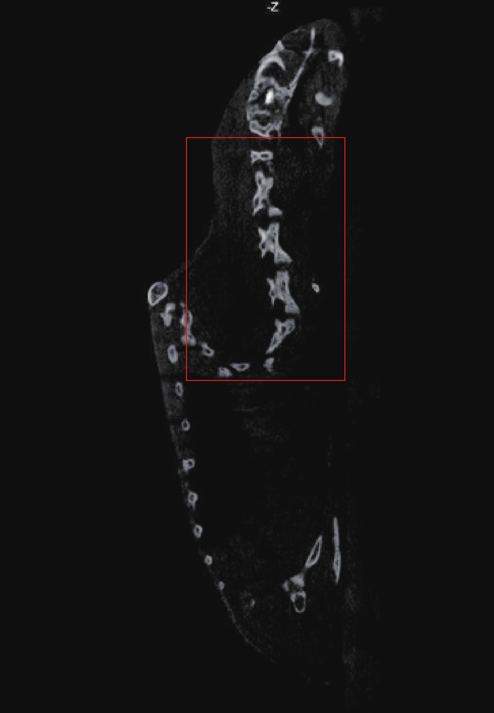
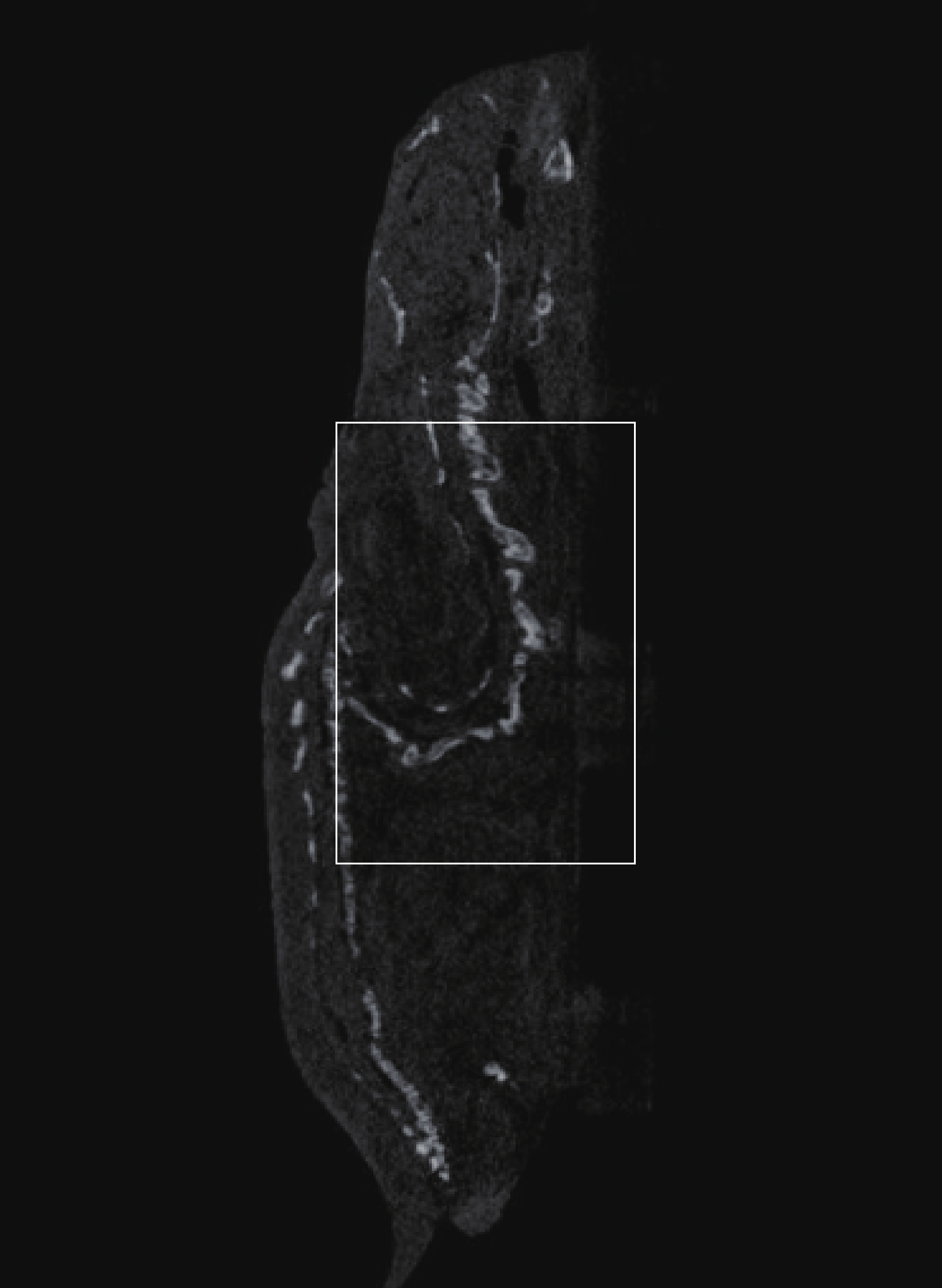
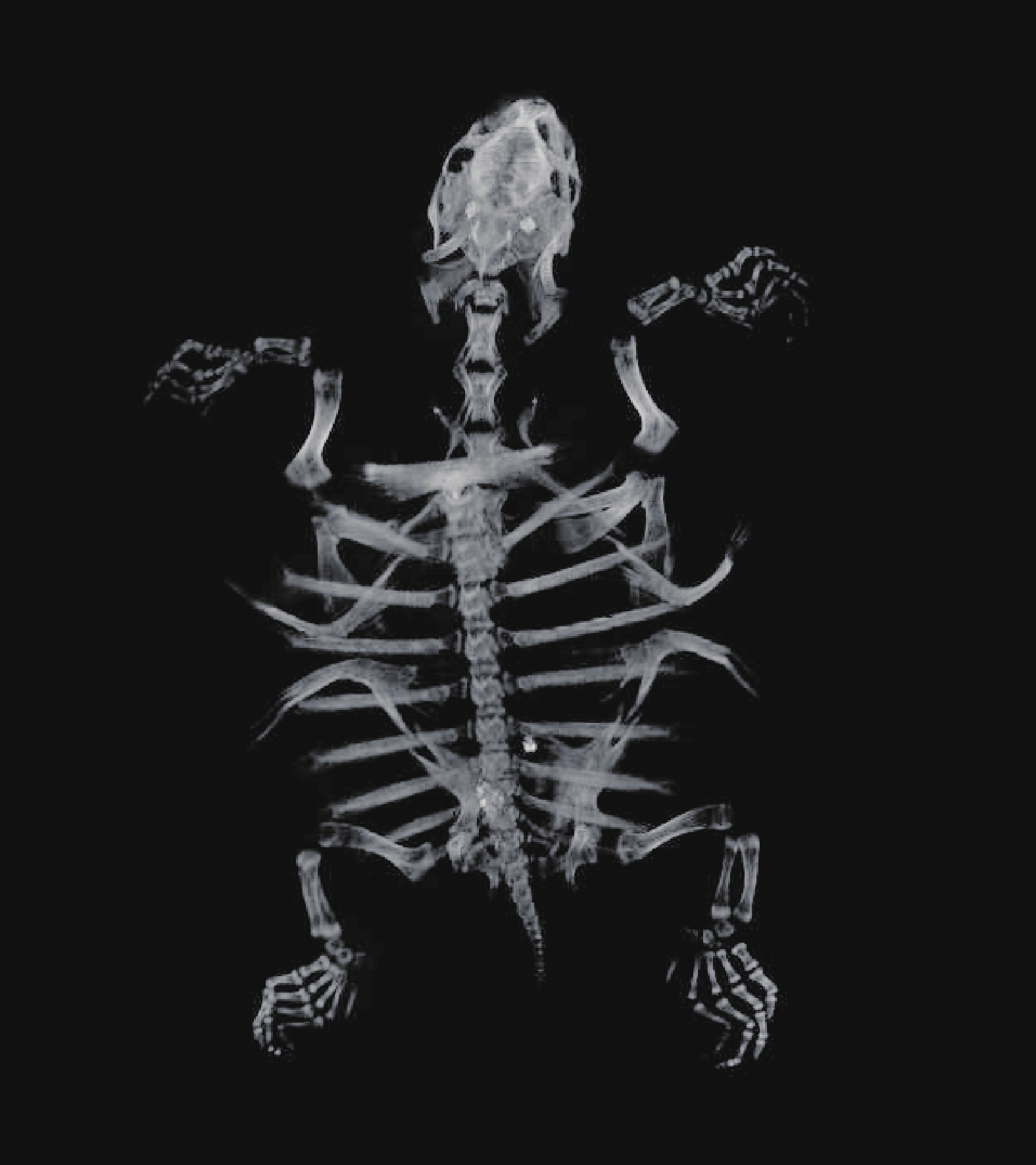
![]() 图 2 鼋脊椎骨侧面CT图(框内示颈椎)Figure 2. Lateral CT image of P. cantorii spine (cervical vertebra shown in frame)
图 2 鼋脊椎骨侧面CT图(框内示颈椎)Figure 2. Lateral CT image of P. cantorii spine (cervical vertebra shown in frame)背甲与肋骨 鼋壳(图 3和图 4)由背甲和腹甲合包而成, 与肋骨(Rib)及脊柱中段愈合, 是鼋赖以生存的保护性构造, 在遇到惊扰时, 头可缩入壳内, 但四肢不能全部缩入。鼋的背甲是一块椭圆形、背面拱起的骨板, 除去皮肤后, 可见由背部骨表面呈虫蚀状纹路, 是由24块小骨板以锯齿状的骨缝缀合而成。最前面一块狭长的横板为颈骨板(Nuchal plate); 正中是一纵列7块方形小骨板, 为椎骨板(Neural plate), 最后一块为三角形。两侧是8对横向长方形的肋骨板(Costal plate), 肋骨板内缘与椎骨板相接, 外缘游离。共有肋骨10对, 第一肋骨较小, 并斜向后方与第二肋骨会合; 第十肋骨也较小, 不甚明显, 仅表现为一突起而不与肋骨板相连。其余的肋骨均附着于各胸椎椎体相接处, 与肋骨板愈合, 远端伸出肋骨板之外。
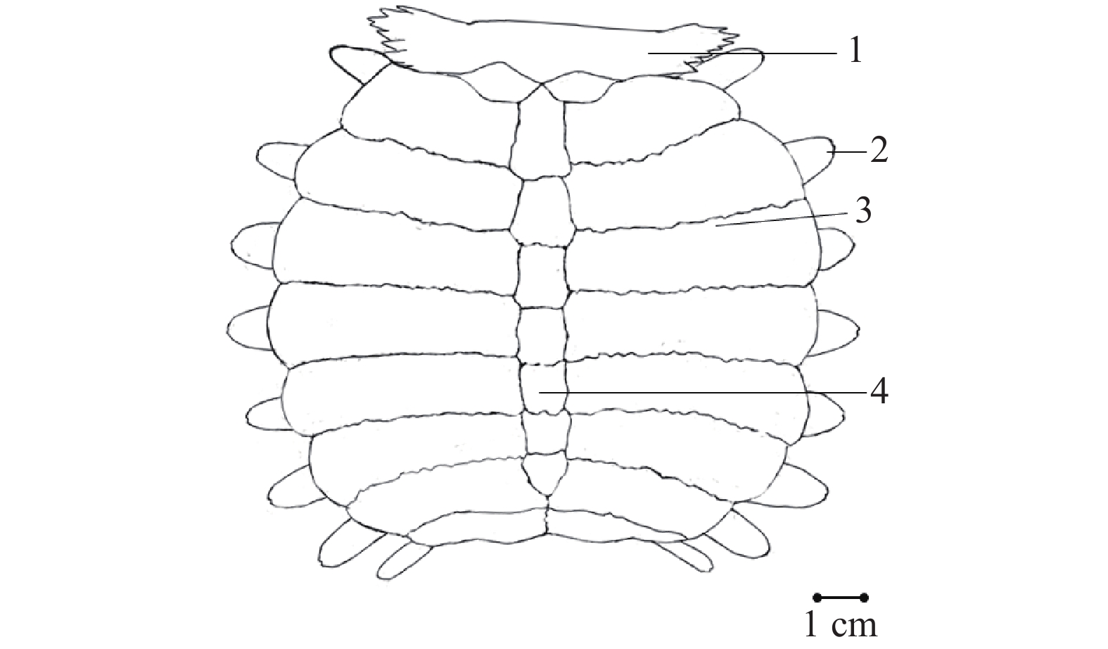
![]() 图 3 鼋背甲背面观1. 颈骨板; 2. 肋骨; 3. 肋骨板; 4. 椎骨板Figure 3. Dorsal view of the carapace of P. cantorii1. Nuchal plate; 2. Rib; 3. Costal plate; 4. Neural plate
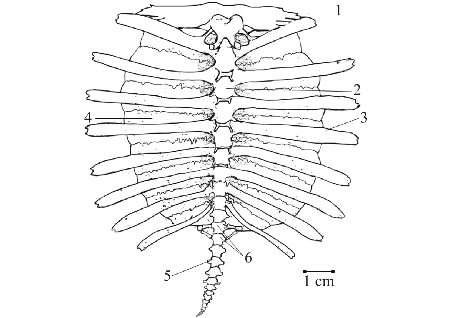
图 3 鼋背甲背面观1. 颈骨板; 2. 肋骨; 3. 肋骨板; 4. 椎骨板Figure 3. Dorsal view of the carapace of P. cantorii1. Nuchal plate; 2. Rib; 3. Costal plate; 4. Neural plate![]() 图 4 鼋背甲腹面观1. 颈骨板; 2. 胸椎; 3. 肋骨; 4. 肋骨板; 5. 尾椎; 6. 荐椎Figure 4. Ventral view of the carapace of P. cantorii1. Nuchal plate; 2. Thoracic vertebra; 3. Rib; 4. Costal plate; 5. Caudal vertebra; 6. Sacral vertebra
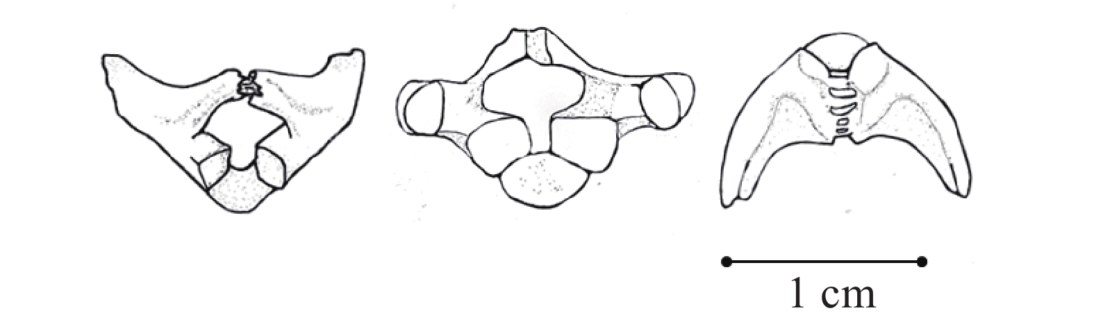
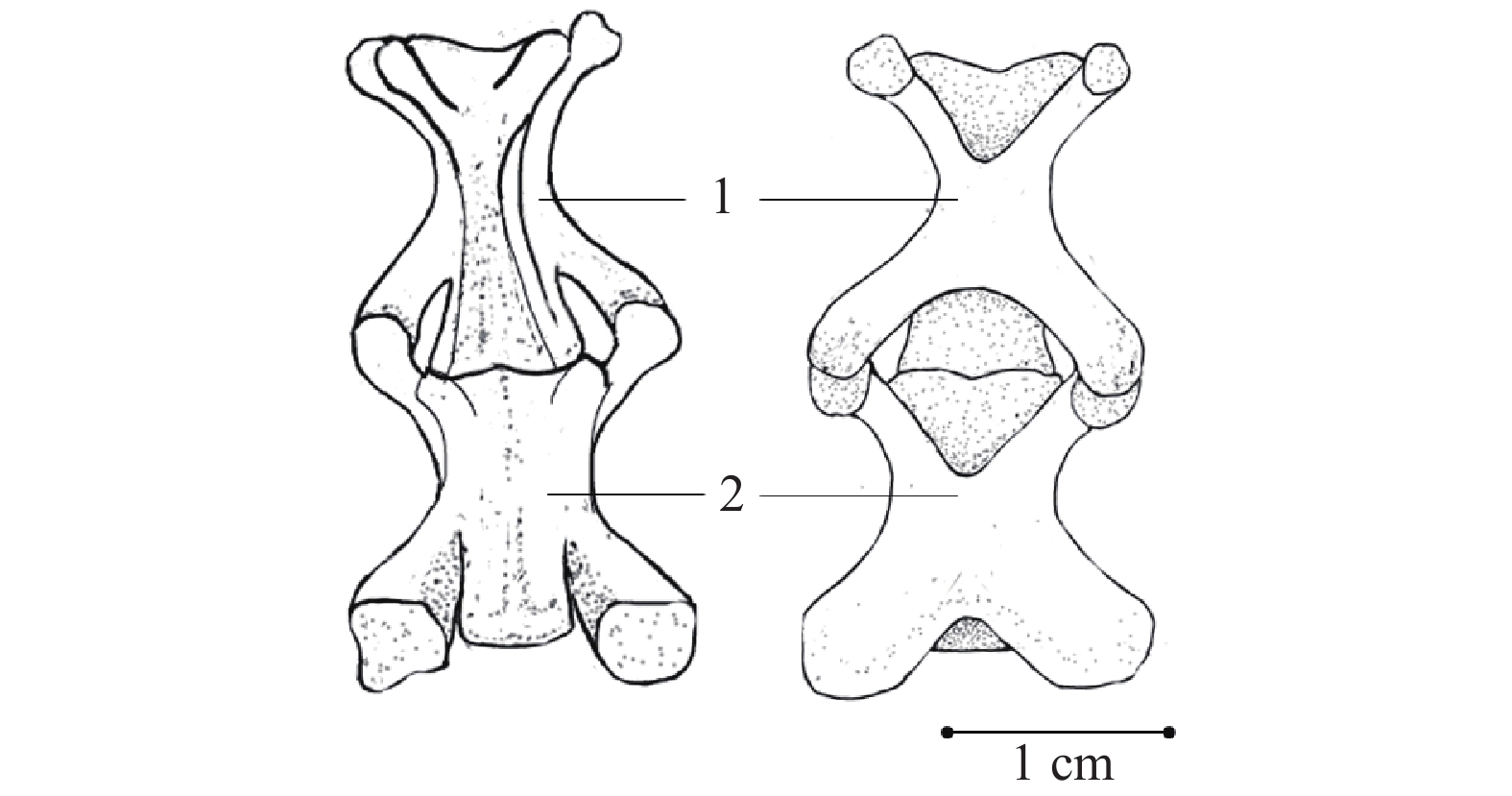
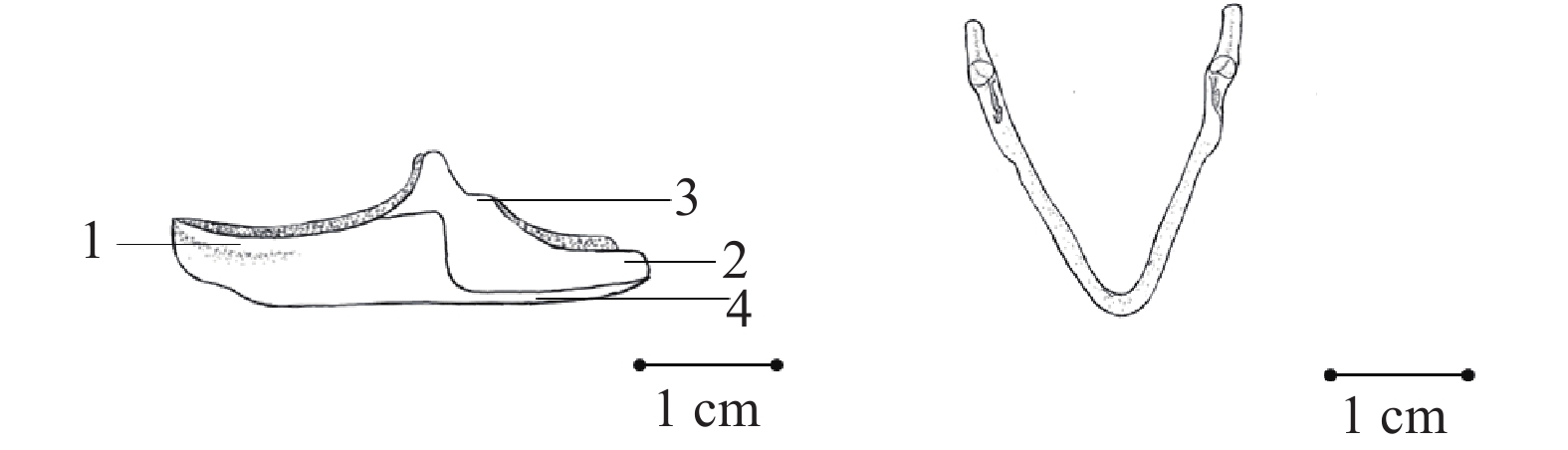
图 4 鼋背甲腹面观1. 颈骨板; 2. 胸椎; 3. 肋骨; 4. 肋骨板; 5. 尾椎; 6. 荐椎Figure 4. Ventral view of the carapace of P. cantorii1. Nuchal plate; 2. Thoracic vertebra; 3. Rib; 4. Costal plate; 5. Caudal vertebra; 6. Sacral vertebra腹甲 腹甲(图 5)相对于背甲更为小而扁平, 只在中段两侧稍向背面弯曲, 以皮肤与背甲相连。除去皮肤后, 可得9块小骨板, 形状互异, 整体呈对称存在。最前面是一对细薄镰刀形状, 前端钝圆的小骨片, 称为新腹骨板(Neoplastron)。其次是一块倒“V”形骨板, 夹角为80°角, 为上腹骨板(Epiplastron)。后面是一对狭长横向的舌腹骨板(Hyoplastron)和一对下腹骨板(Hypoplastron)上下互相愈合。舌腹骨板的外缘与内缘均有齿状突起, 内缘齿状突起数量不一, 且数量常常不对称, 内缘伸向腹甲中线的两个中央腔(Central lacuna), 外缘亦有齿状突起, 突起较少, 弯向背面。下腹板的外缘与内缘也均有刺状突起, 内缘一侧在中央内凹, 凹入处光滑无刺突。最后一对为剑腹骨板(Xiphiplastron), 以内侧的齿状突起嵌合在一起, 而外侧的齿状突起则与下腹骨板内缘的齿状突起相嵌合。
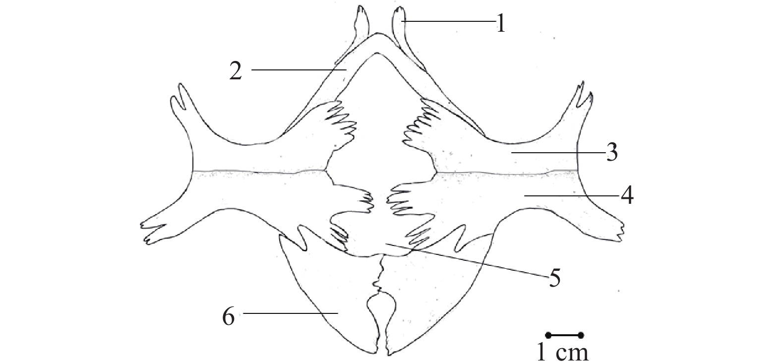
![]() 图 5 鼋腹甲1. 新腹骨板; 2. 上腹骨板; 3. 舌腹骨板; 4. 下腹骨板; 5. 中央腔; 6. 剑腹骨板Figure 5. Plastron of P. cantorii1. Neoplastron; 2. Epiplastron; 3. Hyoplastron; 4. Hypoplastron; 5. Central lacuna; 6. Xiphiplastron
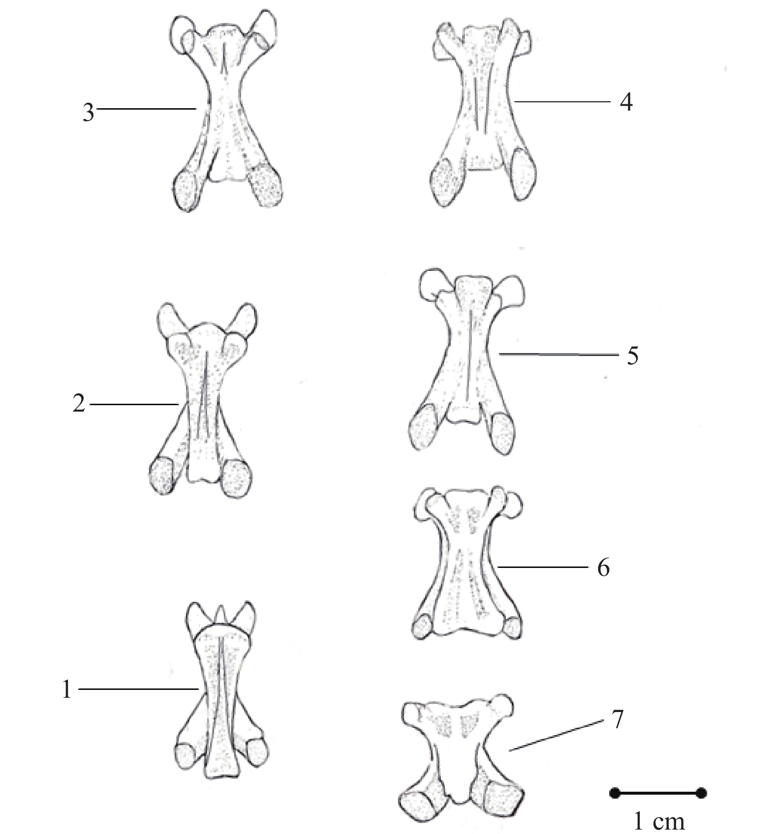
图 5 鼋腹甲1. 新腹骨板; 2. 上腹骨板; 3. 舌腹骨板; 4. 下腹骨板; 5. 中央腔; 6. 剑腹骨板Figure 5. Plastron of P. cantorii1. Neoplastron; 2. Epiplastron; 3. Hyoplastron; 4. Hypoplastron; 5. Central lacuna; 6. Xiphiplastron脊柱 脊柱由颈椎(Cervical vertebra)、胸椎(Thoracic vertebra)、荐椎(Sacral vertebra)和尾椎(Caudal vertebra)构成。颈椎共9枚, 所有颈椎均游离, 不与背甲愈合, 无颈肋, 前后以骨缝相接。第一、二枚颈椎特化为寰椎(Atlas; 图 6)和枢椎(Axis; 图 7)。寰椎呈环状, 由腹面小而成弧形的椎体与背面大的髓弓(Neural arch)共同合围成一个大腔, 中间由一横韧带分隔成腹面的寰椎孔和背面的椎管, 髓弓向后伸出一对较大的后关节突(Postzygapophysis), 与第三颈椎(The third cervical vertebrae)的前关节突(Prezygapophysis)连接。枢椎(图 7)是长方形骨块, 较小, 无椎弓, 前端延展成两个关节突向前伸出, 与寰椎髓弓内侧面相关接; 两关节突的中间伸入寰椎孔; 椎体后端则形成关节凹面, 与第三颈椎的椎体相接。自第三颈椎开始, 为一般典型的颈椎。第三颈椎至第九颈椎(The ninth cervical vertebrae)均由髓体、髓弓及前后关节突构成, 前后关节突分别向前后伸出, 呈八字状, 且每节脊椎的横突(Transverse process)渐渐明显。第三颈椎背面椎管向前伸出呈“m”形, 而第四至第九椎体均向内凹入, 呈“V”形。脊椎背面的髓弓以第五至第七节(图 8和图 9)最为明显, 依次高耸, 到第七节高度尤甚, 第八、第九节(图 10)则趋于平缓, 在第九节几乎呈平滑表面。椎体腹面的纵脊明显, 逐渐低平, 到第八、第九颈椎反而向内凹入。
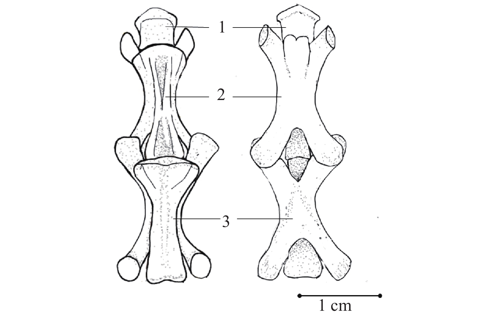
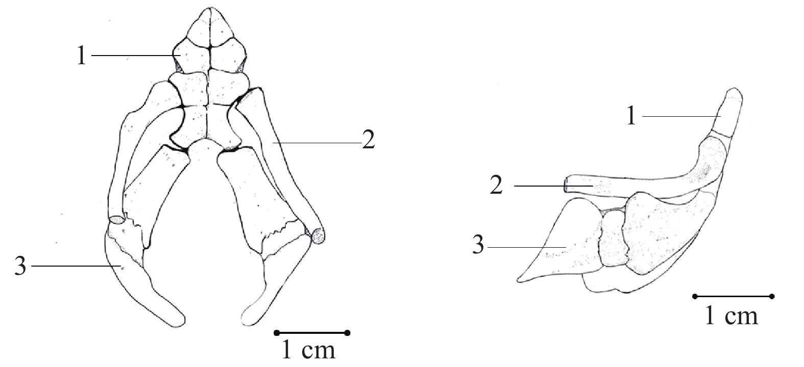
![]() 图 7 鼋第二—四颈椎(左腹面观, 右背面观)1. 枢椎; 2. 第三颈椎; 3. 第四颈椎Figure 7. The second to the fourth cervical vertebrae of P. cantorii (Ventral view on the left, dorsal view on the right)1. Axis; 2. the third cervical spine; 3. the fourth cervical spine
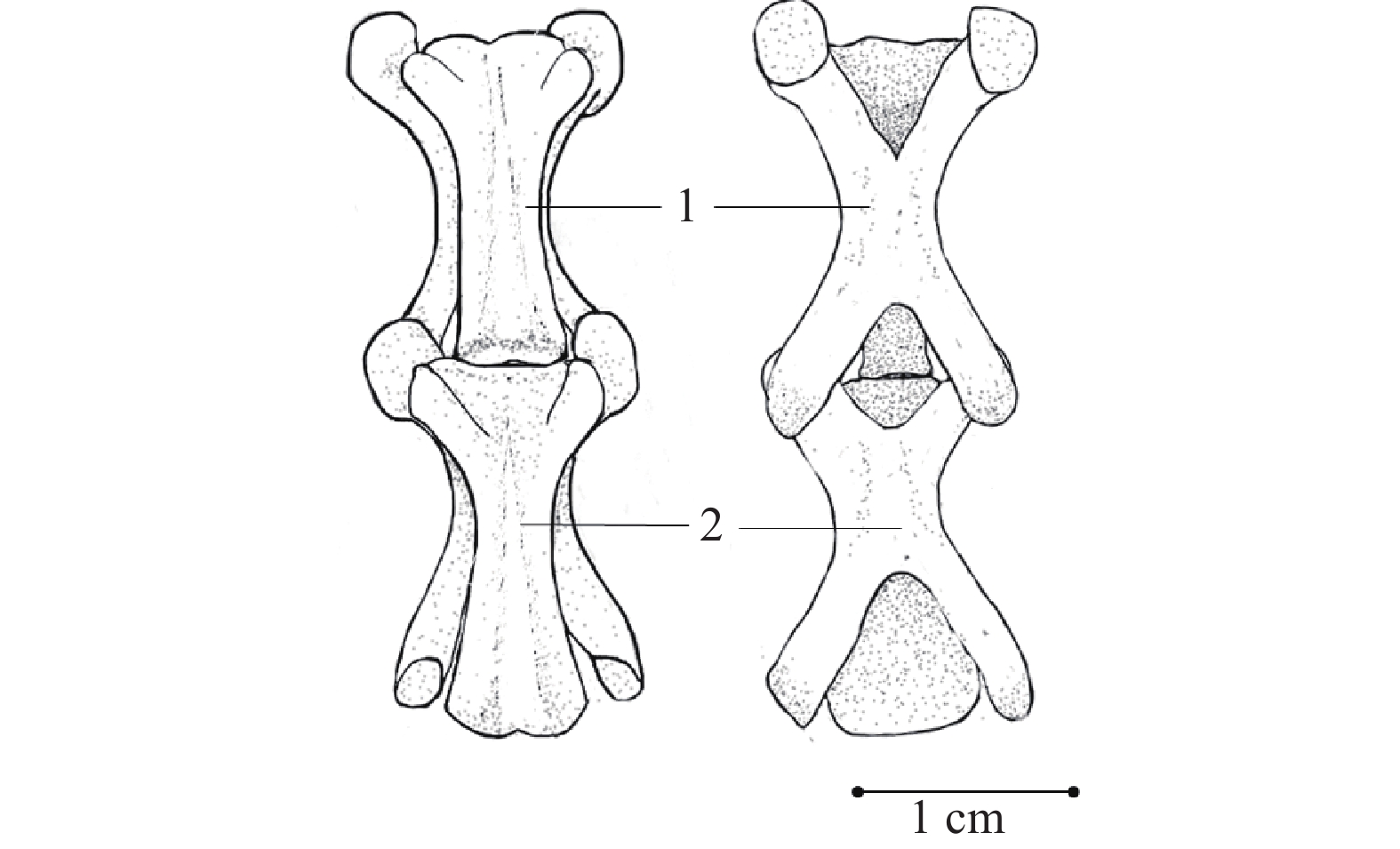
图 7 鼋第二—四颈椎(左腹面观, 右背面观)1. 枢椎; 2. 第三颈椎; 3. 第四颈椎Figure 7. The second to the fourth cervical vertebrae of P. cantorii (Ventral view on the left, dorsal view on the right)1. Axis; 2. the third cervical spine; 3. the fourth cervical spine![]() 图 8 鼋第五—六颈椎(左腹面观, 右背面观)1. 第五颈椎; 2.第六颈椎Figure 8. The fifth to the sixth cervical vertebrae of P. cantorii Ⅱ (Ventral view on the left, dorsal view on the right)1. the fifth cervical vertebra; 2. the sixth cervical vertebra
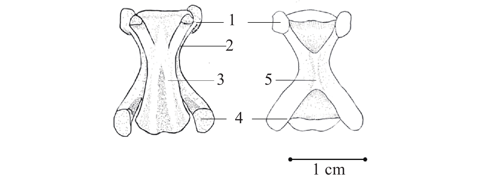
图 8 鼋第五—六颈椎(左腹面观, 右背面观)1. 第五颈椎; 2.第六颈椎Figure 8. The fifth to the sixth cervical vertebrae of P. cantorii Ⅱ (Ventral view on the left, dorsal view on the right)1. the fifth cervical vertebra; 2. the sixth cervical vertebra![]() 图 9 鼋第七颈椎(左腹面观, 右背面观)1. 前关节突; 2. 横突; 3. 棘突; 4. 后关节突; 5. 髓弓Figure 9. The seventh cervical vertebrae of P. cantorii (Ventral view on the left, dorsal view on the right)1. Prezygapophysis; 2. Transverse process; 3. Spinous process; 4. Postzygapophysis; 5. Neural arch
图 9 鼋第七颈椎(左腹面观, 右背面观)1. 前关节突; 2. 横突; 3. 棘突; 4. 后关节突; 5. 髓弓Figure 9. The seventh cervical vertebrae of P. cantorii (Ventral view on the left, dorsal view on the right)1. Prezygapophysis; 2. Transverse process; 3. Spinous process; 4. Postzygapophysis; 5. Neural arch![]() 图 10 鼋第八—九颈椎(左腹面观, 右背面观)1. 第八颈椎 2. 第九颈椎Figure 10. The eighth to ninth cervical vertebrae of P. cantorii (Ventral view on the left, dorsal view on the right)1. the eighth cervical vertebra; 2. the ninth cervical vertebra
图 10 鼋第八—九颈椎(左腹面观, 右背面观)1. 第八颈椎 2. 第九颈椎Figure 10. The eighth to ninth cervical vertebrae of P. cantorii (Ventral view on the left, dorsal view on the right)1. the eighth cervical vertebra; 2. the ninth cervical vertebra胸椎共10枚(图 4)。第一胸椎较为特化, 不与背甲的颈骨板愈合, 前关节突很大, 与第九颈椎的后关节突相接, 前关节突后面伸出一对短小的肋骨附着于第二肋骨上端。从第二到第九胸椎依次变小, 髓弓的背侧部分与背甲椎骨板愈合。胸椎的髓弓与髓弓之间有椎间孔。
荐椎2枚(图 4), 较小, 除前、后关节突外, 并有横突, 上面附着较大的荐肋。两荐肋于远端会合。
尾椎21枚(图 4), 前到后逐个变小, 有明显的髓弓、髓棘和关节突。前关节突较大, 后缘扩展如横突。最后4枚, 仅有棒状的椎体。
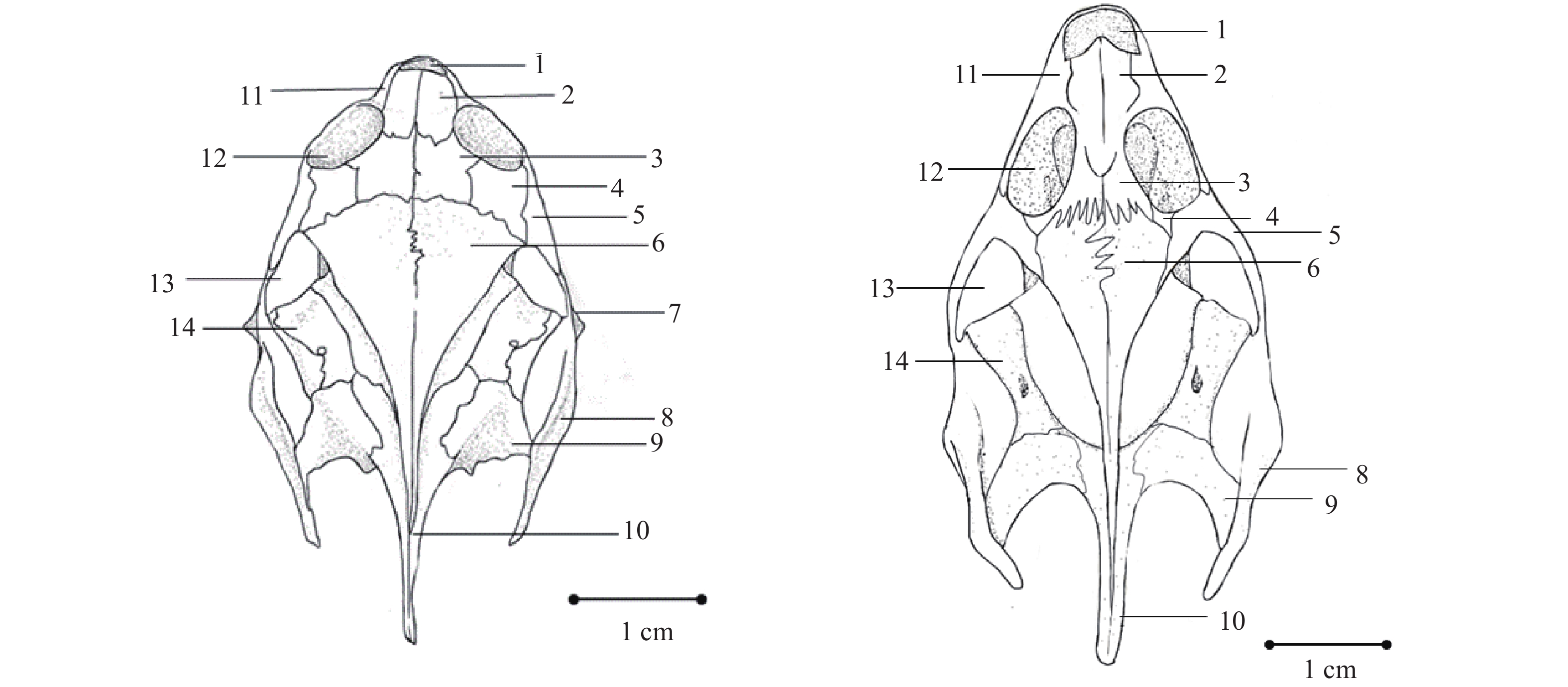
头骨 鼋头骨(图 11—13)除下颌骨外, 各骨块高度愈合, 固定不动, 仅愈合处多具有线状纹。
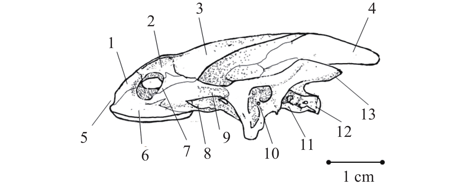
![]() 图 11 鼋头骨侧面观1. 前额骨; 2. 额骨; 3. 顶骨; 4. 上枕骨; 5. 鼻窝; 6. 上颌骨; 7. 后眶骨; 8. 翼骨; 9. 颧骨; 10. 方骨; 11. 基枕骨; 12. 基枕骨髁; 13. 鳞骨Figure 11. Side view of the skull of P. cantorii1. Prefrontal; 2. Frontal; 3. Parictal; 4. Supraoccipital; 5. Fossa olfactorium; 6. Maxilla; 7. Postorbitale; 8. Pterygoid; 9. Zygomaticum; 10. Quadrate; 11. Basioccipital; 12. Condylus basioccipitalis; 13. Squamosal
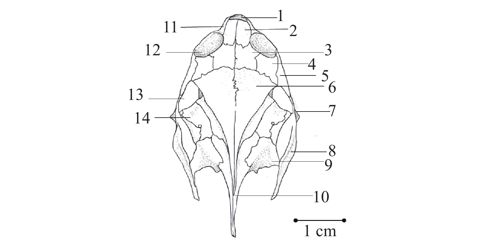
图 11 鼋头骨侧面观1. 前额骨; 2. 额骨; 3. 顶骨; 4. 上枕骨; 5. 鼻窝; 6. 上颌骨; 7. 后眶骨; 8. 翼骨; 9. 颧骨; 10. 方骨; 11. 基枕骨; 12. 基枕骨髁; 13. 鳞骨Figure 11. Side view of the skull of P. cantorii1. Prefrontal; 2. Frontal; 3. Parictal; 4. Supraoccipital; 5. Fossa olfactorium; 6. Maxilla; 7. Postorbitale; 8. Pterygoid; 9. Zygomaticum; 10. Quadrate; 11. Basioccipital; 12. Condylus basioccipitalis; 13. Squamosal![]() 图 12 鼋头骨背面观1. 鼻窝; 2. 前额骨; 3. 额骨; 4. 后眶骨; 5. 颧骨; 6. 顶骨; 7. 方轭骨; 8. 鳞骨; 9. 后耳骨; 10. 上枕骨; 11. 上颌骨; 12. 眼窝; 13. 颞区; 14. 前耳骨Figure 12. View from the back of the skul of P. cantorii1. Fossa olfactorium; 2. Prefrontal; 3. Frontal; 4. Postorbitale; 5. Zygomaticum; 6. Parictal; 7. Quadratojugal; 8. Squamosal; 9. Opisthotic; 10. Supraoccipital; 11. Maxilla; 12. Foramen orbitale; 13. Temporal region; 14. Prootic
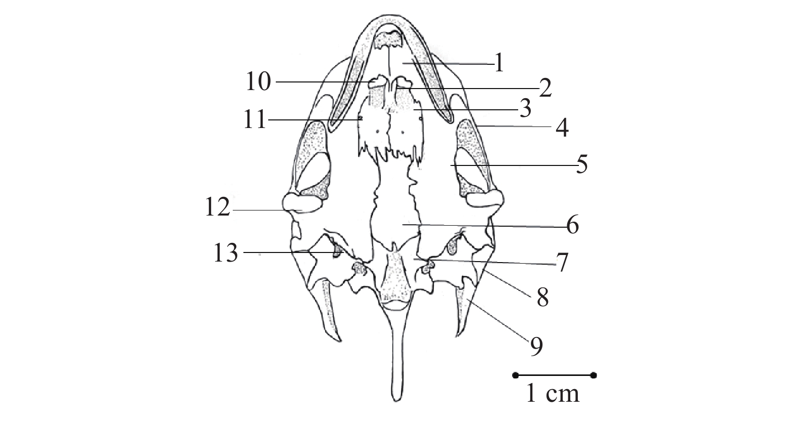
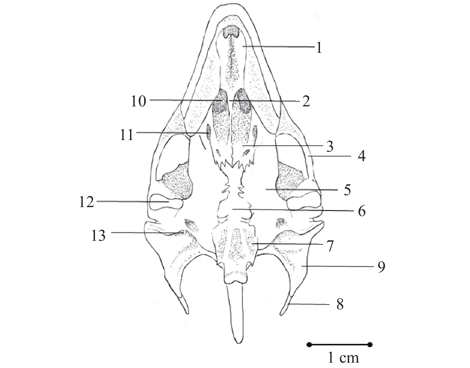
图 12 鼋头骨背面观1. 鼻窝; 2. 前额骨; 3. 额骨; 4. 后眶骨; 5. 颧骨; 6. 顶骨; 7. 方轭骨; 8. 鳞骨; 9. 后耳骨; 10. 上枕骨; 11. 上颌骨; 12. 眼窝; 13. 颞区; 14. 前耳骨Figure 12. View from the back of the skul of P. cantorii1. Fossa olfactorium; 2. Prefrontal; 3. Frontal; 4. Postorbitale; 5. Zygomaticum; 6. Parictal; 7. Quadratojugal; 8. Squamosal; 9. Opisthotic; 10. Supraoccipital; 11. Maxilla; 12. Foramen orbitale; 13. Temporal region; 14. Prootic![]() 图 13 鼋头骨腹面观1. 上颌骨腭突; 2. 犁骨; 3. 腭骨; 4. 颧骨; 5. 翼骨; 6. 基蝶骨; 7. 基枕骨; 8. 鳞骨; 9. 后耳骨; 10. 内鼻孔; 11.腭后孔; 12. 关节; 13. 后破裂孔Figure 13. Ventral view of the skull of P. cantorii1. Palatine process of maxilla; 2. Vomer; 3. Palatinum; 4. Zygomaticum; 5. Pterygoid; 6. Basisphenoid; 7. Basioccipital; 8. Squamosal; 9. Opisthotic; 10. Internal nare; 11. Foramen palatine aborale; 12. Arthrosis; 13. Foramen lacerurm posterius
图 13 鼋头骨腹面观1. 上颌骨腭突; 2. 犁骨; 3. 腭骨; 4. 颧骨; 5. 翼骨; 6. 基蝶骨; 7. 基枕骨; 8. 鳞骨; 9. 后耳骨; 10. 内鼻孔; 11.腭后孔; 12. 关节; 13. 后破裂孔Figure 13. Ventral view of the skull of P. cantorii1. Palatine process of maxilla; 2. Vomer; 3. Palatinum; 4. Zygomaticum; 5. Pterygoid; 6. Basisphenoid; 7. Basioccipital; 8. Squamosal; 9. Opisthotic; 10. Internal nare; 11. Foramen palatine aborale; 12. Arthrosis; 13. Foramen lacerurm posterius(1)上颌骨(Maxilla): 一对, 在头部前端两侧, 较长的底边构成颌缘; 旁侧依着前额骨, 是鼻窝的侧壁; 后端则形成眼窝的前壁, 并与后面的颧骨相接。
(2)前额骨(Prefrontal): 一对, 位于头顶的前方, 形状趋近于长方形。其前端构成鼻窝的后壁; 后端连接额骨; 前缘接上颌骨, 后缘则与额骨共同形成眼窝的内侧壁。
(3)额骨(Frontal): 一对, 在前额骨的后方, 比前额骨更大。前端与前额骨相连; 后端则接顶骨, 外侧接后眶骨。
(4)顶骨(Parictal): 一对, 呈三角形, 在头顶的正中、额骨的后方。前接额骨和后眶骨; 斜边成棱线, 并向下成坡延展, 与前耳骨相接; 后侧则在头顶中线向后成又长又细的骨鞘套在上枕骨的枕骨脊上。
(5)后眶骨(Postorbitale): 一对, 位于额骨的外侧。其前缘构成了鼋眼眶后的壁, 外侧则与颧骨相连。
(6)颧骨(Zygomaticum): 一对, 位于后眶骨的外侧。前与上颌骨相连, 侧与后眶骨相接, 后方则连接方轭骨, 从内表面还伸出内支接着翼骨的内表面。
(7)方轭骨(Quadratojugal): 一对, 细而小, 三叉形。前支伸入颧骨, 旁支伸到鳞骨和前耳骨之间, 后支附在方骨的背缘鼓室壁的上方。`
(8)前耳骨(Prootic): 一对, 在顶骨外侧, 部分被顶骨坡所覆盖, 前缘游离, 后缘接后耳骨。
(9)后耳骨(Opisthotic): 一对, 前支接前耳骨及上枕骨, 后支大部分游离, 波浪形, 小部分与鳞骨后端部分相嵌合。
(10)上枕骨(Supraoccipital): 一块, 长形, 前端插入顶骨, 后端伸出一个剑状的枕骨脊, 脊的一部分被顶骨向后延伸的骨鞘所包裹; 外侧靠后耳骨; 而在枕脊的基部, 有两个突出的部分, 与基枕骨相接共同围成枕骨大孔。
(11)方骨(Quadratum): 一对, 较大略呈方形, 底部中央有一根细小的镫骨; 内侧靠前耳骨和后耳骨; 后端为鳞骨所覆盖, 且后方有明显的关节突与下颌相关接。
(12)鳞骨(Squamosal): 一对, 呈角状, 角尖斜向后延伸。基部位于方骨后端, 并与前耳骨及后耳骨相邻。
(13)上颌骨腭突(Palatine process of maxilla): 一对, 是上腭向口腔顶部水平延展的骨片, 左右相接, 构成次生性的骨质腭。
(14)腭骨(Palatinum): 一对, 薄片状, 在头骨腹面, 基蝶骨的前方, 其前端和上颌骨腭突相接。两片腭骨中间夹着一块小型的犁骨。
(15)犁骨(Vomer): 一块, 细小, 在两腭骨中间。
(16)翼骨(Pterygoid): 一对, 长形, 位于基蝶骨和腭骨的外侧。前端伸向上颌骨腭突和腭骨之间; 后端则伸向方骨与基枕骨之间。
(17)基蝶骨(Basisphenoid): 一块, 位在二翼骨之间, 基枕的前方。基部有短的突起和基枕骨相接。侧部紧挨翼骨, 前缘与腭骨相接。
(18)基枕骨(Basioccipital): 一块, 较小, 棒状, 末端有三叶形的基枕裸, 与枢椎相关接。
(19)下颌骨(Mandible)(图 14): 呈“V”形, 夹角约为50°。为齿骨(Dentary), 隅骨(Angular), 及上隅骨(Surangula)所构成, 隅骨中还包埋着关节骨(Articular), 与方骨的关节突形成下颌关节。
![]() 图 14 鼋下颌骨1. 齿骨; 2. 上隅骨; 3. 关节骨; 4. 隅骨Figure 14. Mandible of P. cantorii1. Dentary; 2. Surangula; 3. Articular; 4. Angular
图 14 鼋下颌骨1. 齿骨; 2. 上隅骨; 3. 关节骨; 4. 隅骨Figure 14. Mandible of P. cantorii1. Dentary; 2. Surangula; 3. Articular; 4. Angular(20)舌骨器(Apparatus hyoideus; 图 15): 位于头骨腹面, 由舌内软骨(Cartilage entoglossalis)(丢失)、 舌骨角(Body of hyoid)、 前角(Cornu anterior)和后角(Cornu posteriors)构成。舌骨体由四对对称的小骨组成。第3对小骨外侧处, 附着一对弯曲的弓形骨为前角。后端还附着一对由3个骨节组成的后角。前角、后角均由腹面弯向背面, 保护着喉头和气管。
![]() 图 15 鼋舌骨1. 舌骨体; 2. 前角; 3. 后角Figure 15. Apparatus hyoideus of P. cantorii1. Body of hyoid; 2. Cornu anterior; 3. Cornu posteriors
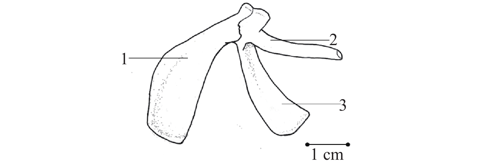
图 15 鼋舌骨1. 舌骨体; 2. 前角; 3. 后角Figure 15. Apparatus hyoideus of P. cantorii1. Body of hyoid; 2. Cornu anterior; 3. Cornu posteriors肩带 由肩胛骨(Scapula)和乌喙骨(Coracoideum)组成(图 16), 肩胛骨有分支伸到腹面为前肩胛骨(Prescapular)。肩胛骨远端以韧带连附在背甲内表面第一对肋骨附近的肋骨板上, 是肩带的背面部分。腹面是前肩胛骨和乌喙骨, 乌喙骨扁平状, 外侧的柄与肩胛骨相关形成肩臼。
![]() 图 16 鼋肩带1. 肩胛骨; 2. 前肩胛骨; 3. 乌喙骨Figure 16. Pectoral girdle of P. cantorii1. Scapula; 2. Prescapular; 3. Coracoideum
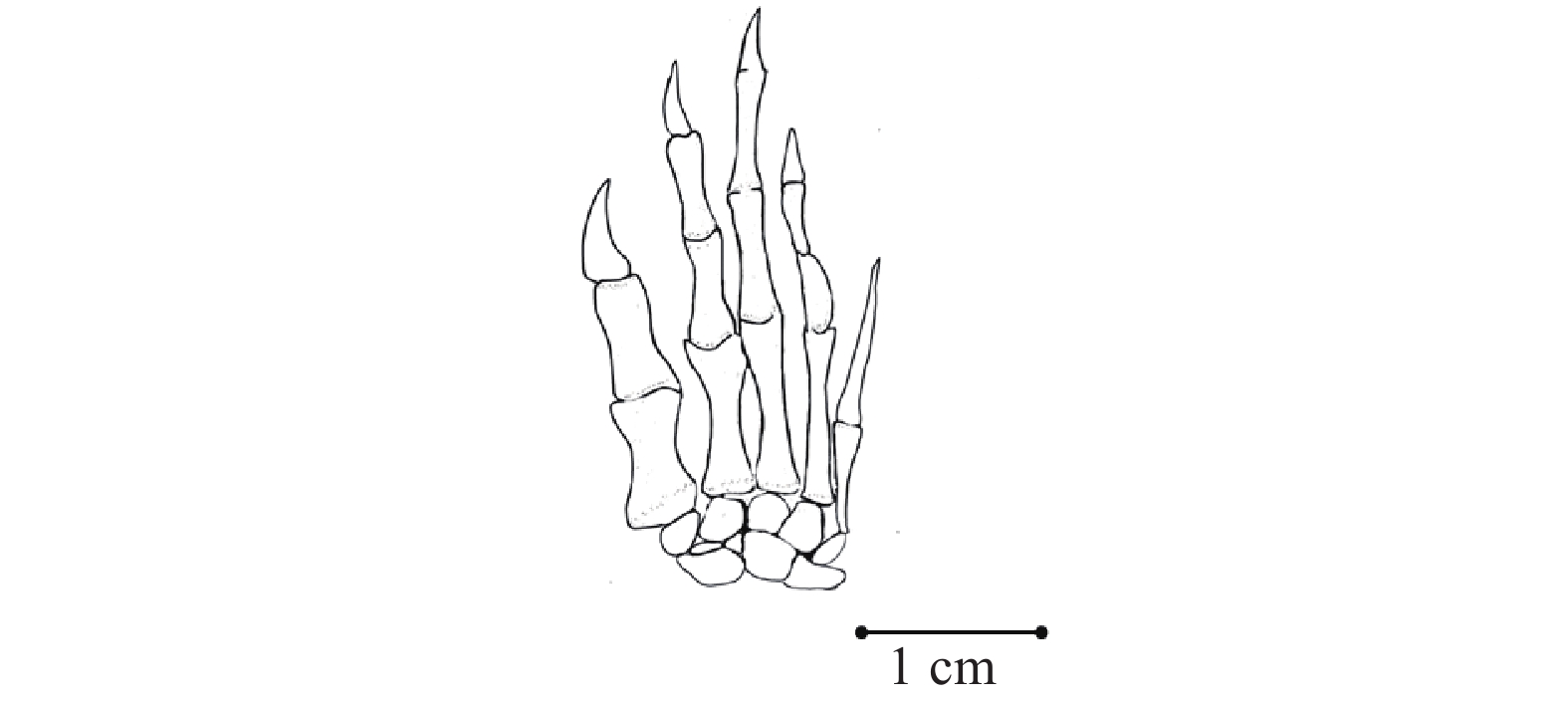
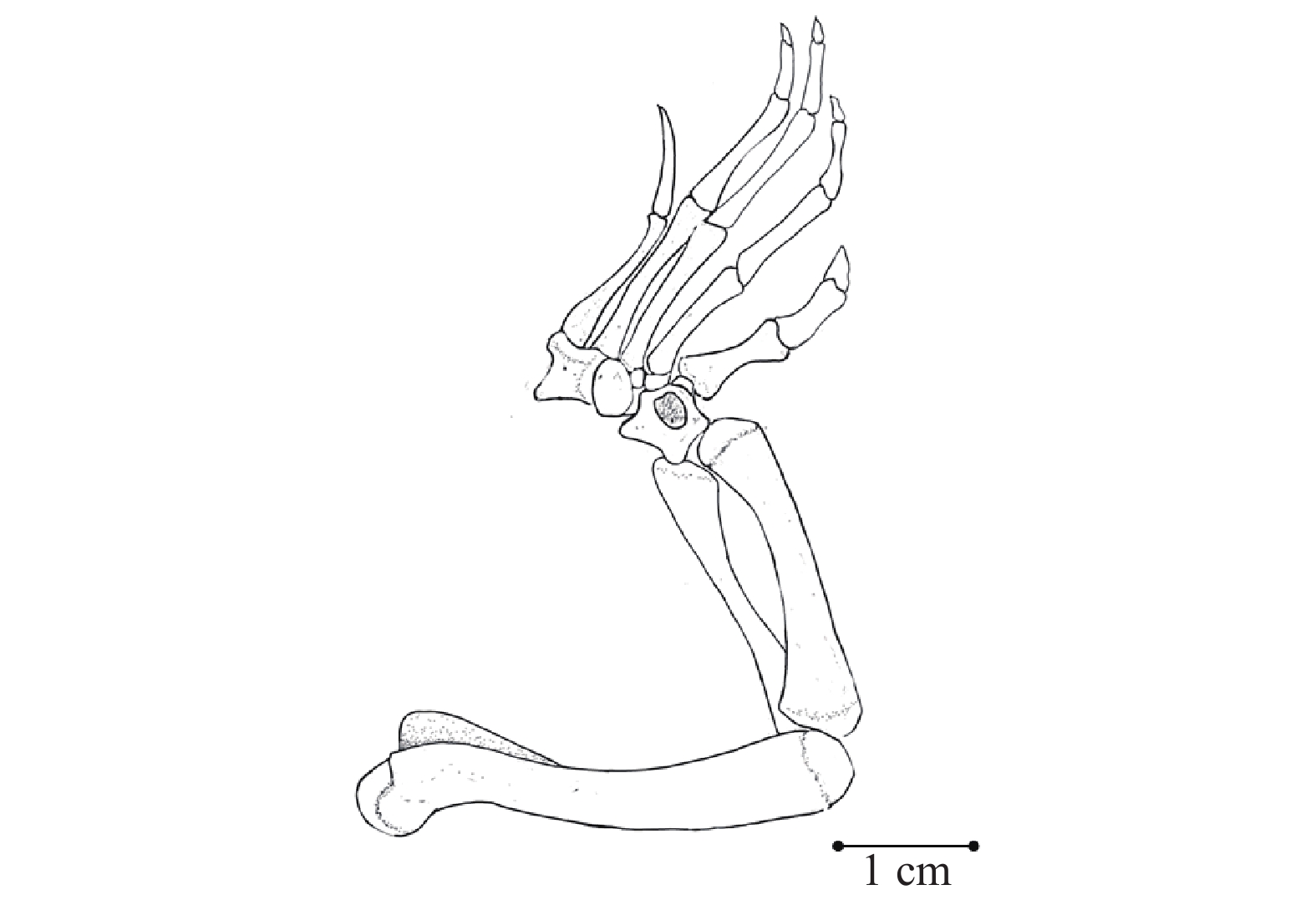
图 16 鼋肩带1. 肩胛骨; 2. 前肩胛骨; 3. 乌喙骨Figure 16. Pectoral girdle of P. cantorii1. Scapula; 2. Prescapular; 3. Coracoideum前肢骨 由肱骨(Humerus)、桡骨(Radius)及尺骨(Ulna)、腕骨(Carpus)、掌骨(Metacarpale)、指骨(Phalanx;图 18—20)等部分组成。肱骨粗短, 弯曲, 近端有球形的肱骨头, 伸入肩臼, 形成可转动的关节。肱骨头旁有两个隆起, 即大结节和小结节, 中间有深沟。远端略扁, 有关节髁与桡骨及尺骨相接。桡骨较细, 尺骨较粗, 两骨并列, 在前端与后端相接, 中间形成空隙。腕骨呈不规则的颗粒状, 互相嵌合, 并与前面的桡、尺骨以及后面的掌骨相接。腕骨共10块, 包括与掌骨一一对应连接的5枚, 以及靠桡骨侧的桡侧腕骨, 靠尺骨侧的尺侧腕骨, 中间部分为间腕骨, 还有两块紧挨在一起的中央腕骨。掌骨五块, 较粗短, 其后的指骨依次细长, 由第一至第五指节各排指骨数依次为2、3、3、3、1。
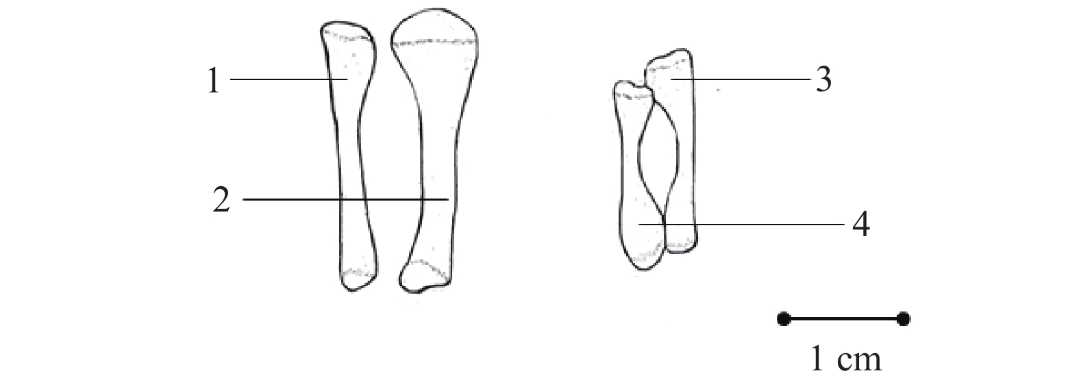
![]() 图 18 部分附肢骨1. 胫骨; 2. 腓骨; 3. 桡骨; 4. 尺骨Figure 18. Partial appendage bone1. Tibia; 2. Fibula; 3. Radius; 4. Ulna
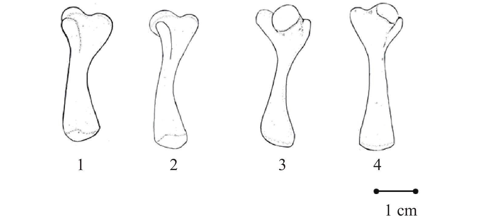
图 18 部分附肢骨1. 胫骨; 2. 腓骨; 3. 桡骨; 4. 尺骨Figure 18. Partial appendage bone1. Tibia; 2. Fibula; 3. Radius; 4. Ulna![]() 图 19 鼋的股骨及肱骨(1、3为股骨; 2、4为肱骨)Figure 19. Femur and humerus of P. cantorii (1, 3 are femur; 2 and 4 are humerus)
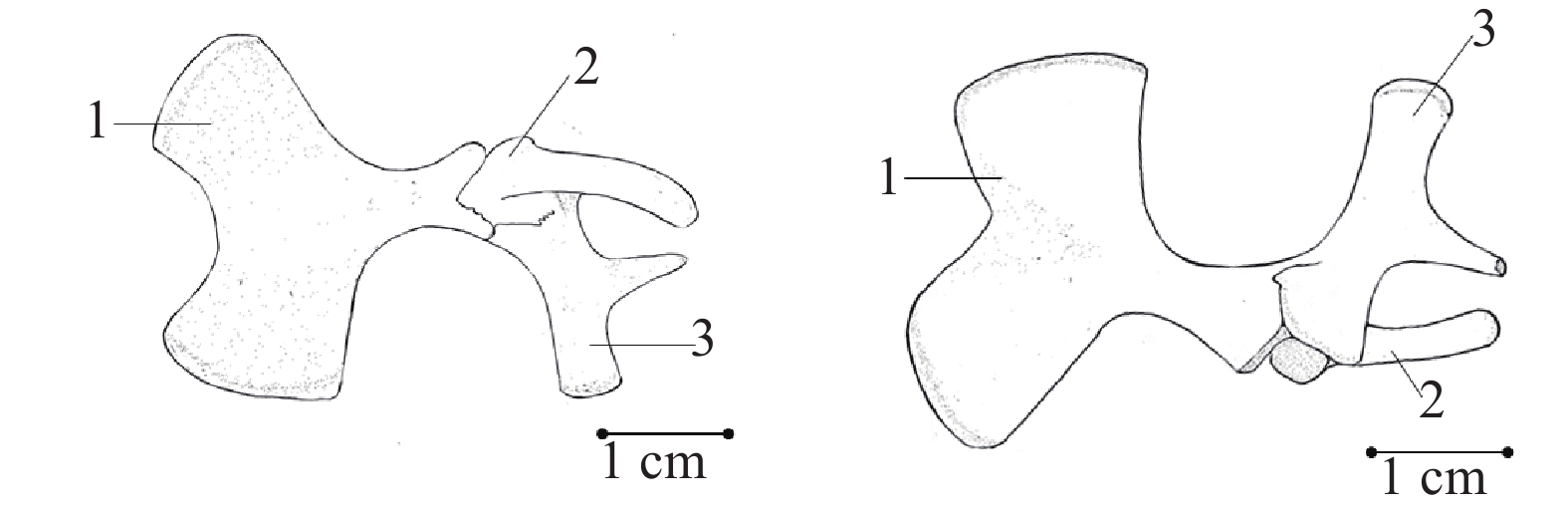
图 19 鼋的股骨及肱骨(1、3为股骨; 2、4为肱骨)Figure 19. Femur and humerus of P. cantorii (1, 3 are femur; 2 and 4 are humerus)腰带 由髂骨(Ilium)、耻骨(Pubis)及坐骨(Ischium)形成(图 17), 三块骨在一端相连而不紧密相接。髂骨较另外两骨更为短细, 柱形, 一端伸向背甲内表面, 以韧带附着于荐助, 另一端与耻骨, 坐骨相连, 其连接处后外侧形成髋臼。耻骨为“Y”形, 较大。坐骨短小, 外侧的上方接耻骨, 中部有一明显突起。
![]() 图 17 鼋腰带1. 耻骨; 2. 髂骨; 3. 坐骨Figure 17. Pelvic girdle of P. cantorii1. Pubis; 2. Ilium; 3. Ischium
图 17 鼋腰带1. 耻骨; 2. 髂骨; 3. 坐骨Figure 17. Pelvic girdle of P. cantorii1. Pubis; 2. Ilium; 3. Ischium后肢骨 由股骨(Femur)、胫骨(Tibia)及腓骨(Fibula)、跗骨(Tarsal)、蹠骨(Metatarsus)、趾骨(Phalanx)等五部分组成(图 18—21)。股骨形态如肱骨, 股骨头较大。胫骨及排骨较桡骨及尺骨长。但胫骨比排骨粗。跗骨6块, 前排在胫骨侧有一块多角形骨, 为距骨, 由胫跗骨和间跗骨愈合而成。在腓骨侧的一块较小, 为跟骨(Calcaneus), 是腓附骨及两块中央附骨合并而成。跟骨与距骨中央还有一较大的及后排的3块小跗骨与蹠骨相接。蹠骨5根, 上接趾骨5排, 各排趾数为2、3、3、3、1。
2.2 中华鳖的头骨和颈椎骨
外部形态上鼋与中华鳖最大的区别来自头部图(中华鳖头部见图 22和图 23), 鼋吻突短, 头部更为浑圆。测鼋眼眶前部至吻突最前端的长度与头骨总长度的比为0.082, 而中华鳖为0.570。解剖二者得颈部脊椎发现, 鼋与中华鳖的颈椎骨数目均为9枚, 第一、第二枚颈椎均特化为寰椎与枢椎, 外形无甚区别(图 24)。而中华鳖的第三至第九颈椎的横突要更明显, 第七颈椎的椎体向上显著翘起, 且第九颈椎腹面椎体前端为尖状(图 25)。最明显的差异来自第三颈椎(图 25和图 26)。从整条颈椎上看, 鼋的脊椎长度与其背甲长度之比为0.66, 中华鳖为1.07。其余部分无特别显著的差别。
![]() 图 22 鼋及中华鳖头骨背面观(左图为鼋头骨; 右图为中华鳖头骨)1. 鼻窝; 2. 前额骨; 3. 额骨; 4. 后眶骨; 5. 颧骨; 6. 顶骨; 7. 方轭骨; 8. 鳞骨; 9. 后耳骨; 10. 上枕骨; 11. 上颌骨; 12. 眼窝; 13. 颞区; 14. 前耳骨Figure 22. View of the back of the P. cantorii and P. sinensis skull (left for P. cantorii and right for P. sinensis)1. Fossa olfactorium; 2. Prefrontal; 3. Frontal; 4. Postorbitale; 5. Zygomaticum; 6. Parictal; 7. Quadratojugal; 8. Squamosal; 9. Opisthotic; 10. Supraoccipital; 11. Maxilla; 12. Foramen orbitale; 13. Temporal region; 14. Prootic
图 22 鼋及中华鳖头骨背面观(左图为鼋头骨; 右图为中华鳖头骨)1. 鼻窝; 2. 前额骨; 3. 额骨; 4. 后眶骨; 5. 颧骨; 6. 顶骨; 7. 方轭骨; 8. 鳞骨; 9. 后耳骨; 10. 上枕骨; 11. 上颌骨; 12. 眼窝; 13. 颞区; 14. 前耳骨Figure 22. View of the back of the P. cantorii and P. sinensis skull (left for P. cantorii and right for P. sinensis)1. Fossa olfactorium; 2. Prefrontal; 3. Frontal; 4. Postorbitale; 5. Zygomaticum; 6. Parictal; 7. Quadratojugal; 8. Squamosal; 9. Opisthotic; 10. Supraoccipital; 11. Maxilla; 12. Foramen orbitale; 13. Temporal region; 14. Prootic![]() 图 23 中华鳖头骨腹面观1. 上颌骨腭突; 2. 犁骨; 3. 腭骨; 4. 颧骨; 5. 翼骨; 6. 基蝶骨; 7. 基枕骨; 8. 鳞骨; 9. 后耳骨; 10. 内鼻孔; 11. 后腭孔; 12. 关节; 13. 后破裂孔Figure 23. P. sinensis Ventral view of the skull1. Palatine process of maxilla; 2. Vomer; 3. Palatinum; 4. Zygomaticum; 5. Pterygoid; 6. Basisphenoid; 7. Basioccipital; 8. Squamosal; 9. Opisthotic; 10. Internal nare; 11. Foramen palatine aborale; 12. Arthrosis; 13. Foramen lacerurm posterius
图 23 中华鳖头骨腹面观1. 上颌骨腭突; 2. 犁骨; 3. 腭骨; 4. 颧骨; 5. 翼骨; 6. 基蝶骨; 7. 基枕骨; 8. 鳞骨; 9. 后耳骨; 10. 内鼻孔; 11. 后腭孔; 12. 关节; 13. 后破裂孔Figure 23. P. sinensis Ventral view of the skull1. Palatine process of maxilla; 2. Vomer; 3. Palatinum; 4. Zygomaticum; 5. Pterygoid; 6. Basisphenoid; 7. Basioccipital; 8. Squamosal; 9. Opisthotic; 10. Internal nare; 11. Foramen palatine aborale; 12. Arthrosis; 13. Foramen lacerurm posterius![]() 图 24 中华鳖(ZJ-20211228)脊椎骨CT图(框内示颈椎)Figure 24. Lateral CT image of P. sinensis (ZJ-20211228) spine (cervical vertebra shown in frame)
图 24 中华鳖(ZJ-20211228)脊椎骨CT图(框内示颈椎)Figure 24. Lateral CT image of P. sinensis (ZJ-20211228) spine (cervical vertebra shown in frame)![]() 图 25 中华鳖颈椎(腹面观)1. 第三颈椎; 2. 第四颈椎; 3. 第五颈椎; 4. 第六颈椎; 5. 第七颈椎; 6. 第八颈椎; 7. 第九颈椎Figure 25. Cervical vertebra of P. sinensis (ventral view)1. the third cervical spine; 2. the fourth cervical vertebra; 3. the fifth cervical vertebra; 4. the sixth cervical vertebra; 5. the seventh cervical vertebra; 6. the eighth cervical vertebra; 7. the ninth cervical vertebra
图 25 中华鳖颈椎(腹面观)1. 第三颈椎; 2. 第四颈椎; 3. 第五颈椎; 4. 第六颈椎; 5. 第七颈椎; 6. 第八颈椎; 7. 第九颈椎Figure 25. Cervical vertebra of P. sinensis (ventral view)1. the third cervical spine; 2. the fourth cervical vertebra; 3. the fifth cervical vertebra; 4. the sixth cervical vertebra; 5. the seventh cervical vertebra; 6. the eighth cervical vertebra; 7. the ninth cervical vertebra![]() 图 26 中华鳖第三颈椎背面观Figure 26. View of the dorsal side of the third cervical spine of P. sinensis
图 26 中华鳖第三颈椎背面观Figure 26. View of the dorsal side of the third cervical spine of P. sinensis3. 讨论
3.1 鼋与中华鳖的骨骼与其他龟类骨骼的比较
本实验中所解剖的鼋个体是2只病死的鼋个体, 均未达到性成熟。在《中国动物志》中, 张孟闻[12]等对6个20—70 kg的性成熟鼋个体标本进行研究, 得到了一系列数据描述。相比之下, 可见幼体鼋与《中国动物志》中所描述性成熟个体骨骼除体积外无甚差异。而鼋与中华鳖腹甲中的新腹骨板是其他龟类, 诸如海龟[14](Chelonia mydas)与四爪陆龟[8]中所不存在的结构, 推测此为龟与鳖在骨骼系统中的一大区别。同时, 鼋与中华鳖的腹甲中没有其他龟类所具有的内腹骨板。此外, 鼋与中华鳖具有9枚颈椎骨, 较海龟[14]所具有的8枚更多。
3.2 鼋骨骼系统对于其进化地位及生态适应性的探讨
鳖类动物是爬行动物中十分特殊的一类, 有些学者试图将鳖类另立为一个亚目, 视作潜颈龟类中的奇特类群。鳖类动物是由龟类进化而来, 最早出现在至今1.4亿年之遥的侏罗纪晚期[12], 在漫长的物种演变和进化方面具有重要参考研究价值[4]。在对于爬行动物的长期研究中, 学者们一致认为, 爬行动物颅骨上颞窝的特征是一个十分可行的分类依据, 即在爬行动物的演化中, 都呈现了颅骨必须经过一个下颞窝类阶段, 说明各种颞窝类的形成先出现的是无颞窝类; 其次是下颞窝类, 再次是上颞窝的形成, 即具有上下两个颞窝; 然后下颞窝收敛, 只留缝隙。鼋是龟鳖类动物, 无颞窝, 但眼有一凹洼, 其功能可与颞窝等价, 可见其属原始爬行动物, 在动物进化上有着重要的地位。
鳖类动物较为扁平的体型减少了它们在水、沙、泥中运动时的阻力。鼋没有龟类动物甲壳外圈厚重的缘盾, 且腰带也更为轻薄, 整个骨骼质量显著下降, 加上其柔软的裙边组织, 将有助于其在水中的浮力和灵巧程度。另外, 鼋较龟类更大的耻骨和坐骨突都为肌肉附着提供了更多的表面积, 大大提高了其在水中的游泳速度。同时, 鼋及中华鳖的颈椎长度与背甲长度之比和实际观察二者捕食情况相一致。中华鳖头部的活动范围更大, 可对四周相当于其背甲长度距离的目标做出攻击, 利于其捕食及防御外来物的攻击。而鼋颈部伸出的距离仅为背甲的约一半长度, 活动范围则较小。
-
![]()
图 2 鼋脊椎骨侧面CT图(框内示颈椎)
Figure 2. Lateral CT image of P. cantorii spine (cervical vertebra shown in frame)
![]()
图 3 鼋背甲背面观
1. 颈骨板; 2. 肋骨; 3. 肋骨板; 4. 椎骨板
Figure 3. Dorsal view of the carapace of P. cantorii
1. Nuchal plate; 2. Rib; 3. Costal plate; 4. Neural plate
![]()
图 4 鼋背甲腹面观
1. 颈骨板; 2. 胸椎; 3. 肋骨; 4. 肋骨板; 5. 尾椎; 6. 荐椎
Figure 4. Ventral view of the carapace of P. cantorii
1. Nuchal plate; 2. Thoracic vertebra; 3. Rib; 4. Costal plate; 5. Caudal vertebra; 6. Sacral vertebra
![]()
图 5 鼋腹甲
1. 新腹骨板; 2. 上腹骨板; 3. 舌腹骨板; 4. 下腹骨板; 5. 中央腔; 6. 剑腹骨板
Figure 5. Plastron of P. cantorii
1. Neoplastron; 2. Epiplastron; 3. Hyoplastron; 4. Hypoplastron; 5. Central lacuna; 6. Xiphiplastron
![]()
图 7 鼋第二—四颈椎(左腹面观, 右背面观)
1. 枢椎; 2. 第三颈椎; 3. 第四颈椎
Figure 7. The second to the fourth cervical vertebrae of P. cantorii (Ventral view on the left, dorsal view on the right)
1. Axis; 2. the third cervical spine; 3. the fourth cervical spine
![]()
图 8 鼋第五—六颈椎(左腹面观, 右背面观)
1. 第五颈椎; 2.第六颈椎
Figure 8. The fifth to the sixth cervical vertebrae of P. cantorii Ⅱ (Ventral view on the left, dorsal view on the right)
1. the fifth cervical vertebra; 2. the sixth cervical vertebra
![]()
图 9 鼋第七颈椎(左腹面观, 右背面观)
1. 前关节突; 2. 横突; 3. 棘突; 4. 后关节突; 5. 髓弓
Figure 9. The seventh cervical vertebrae of P. cantorii (Ventral view on the left, dorsal view on the right)
1. Prezygapophysis; 2. Transverse process; 3. Spinous process; 4. Postzygapophysis; 5. Neural arch
![]()
图 10 鼋第八—九颈椎(左腹面观, 右背面观)
1. 第八颈椎 2. 第九颈椎
Figure 10. The eighth to ninth cervical vertebrae of P. cantorii (Ventral view on the left, dorsal view on the right)
1. the eighth cervical vertebra; 2. the ninth cervical vertebra
![]()
图 11 鼋头骨侧面观
1. 前额骨; 2. 额骨; 3. 顶骨; 4. 上枕骨; 5. 鼻窝; 6. 上颌骨; 7. 后眶骨; 8. 翼骨; 9. 颧骨; 10. 方骨; 11. 基枕骨; 12. 基枕骨髁; 13. 鳞骨
Figure 11. Side view of the skull of P. cantorii
1. Prefrontal; 2. Frontal; 3. Parictal; 4. Supraoccipital; 5. Fossa olfactorium; 6. Maxilla; 7. Postorbitale; 8. Pterygoid; 9. Zygomaticum; 10. Quadrate; 11. Basioccipital; 12. Condylus basioccipitalis; 13. Squamosal
![]()
图 12 鼋头骨背面观
1. 鼻窝; 2. 前额骨; 3. 额骨; 4. 后眶骨; 5. 颧骨; 6. 顶骨; 7. 方轭骨; 8. 鳞骨; 9. 后耳骨; 10. 上枕骨; 11. 上颌骨; 12. 眼窝; 13. 颞区; 14. 前耳骨
Figure 12. View from the back of the skul of P. cantorii
1. Fossa olfactorium; 2. Prefrontal; 3. Frontal; 4. Postorbitale; 5. Zygomaticum; 6. Parictal; 7. Quadratojugal; 8. Squamosal; 9. Opisthotic; 10. Supraoccipital; 11. Maxilla; 12. Foramen orbitale; 13. Temporal region; 14. Prootic
![]()
图 13 鼋头骨腹面观
1. 上颌骨腭突; 2. 犁骨; 3. 腭骨; 4. 颧骨; 5. 翼骨; 6. 基蝶骨; 7. 基枕骨; 8. 鳞骨; 9. 后耳骨; 10. 内鼻孔; 11.腭后孔; 12. 关节; 13. 后破裂孔
Figure 13. Ventral view of the skull of P. cantorii
1. Palatine process of maxilla; 2. Vomer; 3. Palatinum; 4. Zygomaticum; 5. Pterygoid; 6. Basisphenoid; 7. Basioccipital; 8. Squamosal; 9. Opisthotic; 10. Internal nare; 11. Foramen palatine aborale; 12. Arthrosis; 13. Foramen lacerurm posterius
![]()
图 14 鼋下颌骨
1. 齿骨; 2. 上隅骨; 3. 关节骨; 4. 隅骨
Figure 14. Mandible of P. cantorii
1. Dentary; 2. Surangula; 3. Articular; 4. Angular
![]()
图 15 鼋舌骨
1. 舌骨体; 2. 前角; 3. 后角
Figure 15. Apparatus hyoideus of P. cantorii
1. Body of hyoid; 2. Cornu anterior; 3. Cornu posteriors
![]()
图 16 鼋肩带
1. 肩胛骨; 2. 前肩胛骨; 3. 乌喙骨
Figure 16. Pectoral girdle of P. cantorii
1. Scapula; 2. Prescapular; 3. Coracoideum
![]()
图 18 部分附肢骨
1. 胫骨; 2. 腓骨; 3. 桡骨; 4. 尺骨
Figure 18. Partial appendage bone
1. Tibia; 2. Fibula; 3. Radius; 4. Ulna
![]()
图 19 鼋的股骨及肱骨(1、3为股骨; 2、4为肱骨)
Figure 19. Femur and humerus of P. cantorii (1, 3 are femur; 2 and 4 are humerus)
![]()
图 17 鼋腰带
1. 耻骨; 2. 髂骨; 3. 坐骨
Figure 17. Pelvic girdle of P. cantorii
1. Pubis; 2. Ilium; 3. Ischium
![]()
图 22 鼋及中华鳖头骨背面观(左图为鼋头骨; 右图为中华鳖头骨)
1. 鼻窝; 2. 前额骨; 3. 额骨; 4. 后眶骨; 5. 颧骨; 6. 顶骨; 7. 方轭骨; 8. 鳞骨; 9. 后耳骨; 10. 上枕骨; 11. 上颌骨; 12. 眼窝; 13. 颞区; 14. 前耳骨
Figure 22. View of the back of the P. cantorii and P. sinensis skull (left for P. cantorii and right for P. sinensis)
1. Fossa olfactorium; 2. Prefrontal; 3. Frontal; 4. Postorbitale; 5. Zygomaticum; 6. Parictal; 7. Quadratojugal; 8. Squamosal; 9. Opisthotic; 10. Supraoccipital; 11. Maxilla; 12. Foramen orbitale; 13. Temporal region; 14. Prootic
![]()
图 23 中华鳖头骨腹面观
1. 上颌骨腭突; 2. 犁骨; 3. 腭骨; 4. 颧骨; 5. 翼骨; 6. 基蝶骨; 7. 基枕骨; 8. 鳞骨; 9. 后耳骨; 10. 内鼻孔; 11. 后腭孔; 12. 关节; 13. 后破裂孔
Figure 23. P. sinensis Ventral view of the skull
1. Palatine process of maxilla; 2. Vomer; 3. Palatinum; 4. Zygomaticum; 5. Pterygoid; 6. Basisphenoid; 7. Basioccipital; 8. Squamosal; 9. Opisthotic; 10. Internal nare; 11. Foramen palatine aborale; 12. Arthrosis; 13. Foramen lacerurm posterius
![]()
图 24 中华鳖(ZJ-20211228)脊椎骨CT图(框内示颈椎)
Figure 24. Lateral CT image of P. sinensis (ZJ-20211228) spine (cervical vertebra shown in frame)
![]()
图 25 中华鳖颈椎(腹面观)
1. 第三颈椎; 2. 第四颈椎; 3. 第五颈椎; 4. 第六颈椎; 5. 第七颈椎; 6. 第八颈椎; 7. 第九颈椎
Figure 25. Cervical vertebra of P. sinensis (ventral view)
1. the third cervical spine; 2. the fourth cervical vertebra; 3. the fifth cervical vertebra; 4. the sixth cervical vertebra; 5. the seventh cervical vertebra; 6. the eighth cervical vertebra; 7. the ninth cervical vertebra
-
[1] 彭一良, 李艳杰. 浅析动物骨骼的起源及其在生物进化上的意义 [J]. 黑龙江农垦师专学报, 2002(2): 85-87. Peng Y L, Li Y J. A brief analysis of the origin of animal skeleton and its significance in biological evolution [J]. Journal of Heilongjiang Nongken Normal College, 2002(2): 85-87.
[2] 王震. 13种兽类肱骨的形态学比较 [J]. 安徽农业科学, 2021, 49(4): 85-87. Wang Z. Morphological comparison of humerus among 13 species of mammals [J]. Journal of Anhui Agricultural Sciences, 2021, 49(4): 85-87.
[3] Fritz U, Havas P. Order Testudines: 2013 update [J]. Zootaxa, 2013, 3703(1): 12-14. doi: 10.11646/zootaxa.3703.1.4
[4] 周婷, 李丕鹏. 中国龟鳖物种多样性及濒危现状 [J]. 四川动物, 2007, 26(2): 463-467. doi: 10.3969/j.issn.1000-7083.2007.02.069 Zhou T, Li P P. Chelonian species diversity and current status in China [J]. Sichuan Journal of Zoology, 2007, 26(2): 463-467. doi: 10.3969/j.issn.1000-7083.2007.02.069
[5] Gaffney E S. The side-necked turtle family Chelidae: A theory of relationships using shared derived characters [J]. American Museum Novitates, 1977, 620(2): 1-28.
[6] Melan P A. The phylogenetic relationships of soft-shelled turtles (family Trionychidae) [J]. Bulletin of the American Museum of Natural History, 1987(186): 1-101.
[7] 傅金钟. 中国产龟鳖类分类研究概述 [J]. 动物学杂志, 1993, 28(1): 58-61. Fu J Z. A summary of taxonomic studies on Tesudines in China [J]. Chinese Journal of Zoology, 1993, 28(1): 58-61.
[8] 许设科, 刘志霄. 四爪陆龟骨骼系统的解剖 [J]. 新疆大学学报 (自然科学版), 1993, 10(1): 93-102. Xu S K, Liu Z X. The anatomy of the skeletal of Testudo horsfieldi gray [J]. Journal of Xinjiang University (Natural Science Edition), 1993, 10(1): 93-102
[9] 沈卉君, 虞快. 中华鳖的解剖研究 Ⅰ、骨骼系统 [J]. 上海师范学院学报(自然科学版), 1981(3): 87-100. Shen H J, Yu K. Study on the anatomy of Chinese soft-shell turtle Ⅰ, skeletal system [J]. Journal of Shanghai Normal University (Natural Sciences), 1981(3): 87-100
[10] 吴佑民, 王宇. 浙江瓯江流域鼋的生态观察 [J]. 动物学杂志, 1987, 22(3): 33-36. Wu Y M, Wang Y. Observation on the ecology of Asian giant soft-shelled turtles in Oujiang River basin of Zhejiang Province [J]. Chinese Journal of Zoology, 1987, 22(3): 33-36.
[11] Hong X Y, Cai X D, Chen C, et al. Conservation status of the Asian giant soft-shell turtle (Pelochelys cantorii) in China [J]. Chelonian Conservation and Biology, 2019, 18(1): 68-74. doi: 10.2744/CCB-1365.1
[12] 农业农村部. 农业农村部关于印发《鼋拯救行动计划(2019-2035年)》的通知 [R]. 中华人民共和国农业农村部公报, 2019(12): 29-36. Ministry of Agriculture and Rural Affairs of the People’s Republic of China. Circular of Ministry of Agriculture and Rural Affairs on Issuing Asian Giant Soft-shelled Turtles Rescue Action Plan (2019-2035) [R]. Gazette of the Ministry of Agriculture and Affairs of the People’s Republic of China, 2019(12): 29-36.
[13] 张孟闻, 宗愉, 马积藩, 等. 中国动物志—爬行纲 Ⅰ卷 [M]. 北京: 科学出版社, 1998: 58-60. Zhang M W, Zong Y, Ma J F, et al. Fauna Sinica, ReptileⅠ [M]. Beijing: Science Press, 1998: 58-60.
[14] Wyneken J. 海龟解剖 [M]. 广州: 暨南大学出版社, 2015: 52- 53. Wyneken J. The Anatomy of Sea Turtles [M]. Guangzhou: Jinan University Press, 2015: 52-53.
-
期刊类型引用(4)
1. 王永明,杨木牛,高小平,史晋绒,郭佳萍,谢碧文,李燕平. 2种副沙鳅属鱼类骨骼解剖学比较研究. 四川动物. 2025(01): 57-68 .  百度学术
百度学术
2. 叶锴,吴丞昊,梁谢恩,单程雪,黄天来. 基于物联网技术的鼋自然保护区生态监测系统设计. 绿色科技. 2024(04): 34-37+43 . 百度学术
3. 万刚,曾嘉伟,肖何威,熊钢,靳利忠,王晓清,胡亚洲. 九肋鳖形态及遗传特性分析. 水生生物学报. 2023(09): 1534-1544 . 本站查看
4. 黄保友,楼灵媛,樊嘉伟,孙伟,钱国英,葛楚天,王宗吉. 基于全基因组的龟鳖目Hox基因序列特征及进化分析. 水生生物学报. 2023(10): 1617-1627 . 本站查看
其他类型引用(0)
 下载:
下载:

计量
- 文章访问数: 2173
- HTML全文浏览量: 574
- PDF下载量: 113
- 被引次数: 4